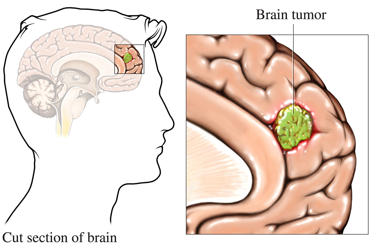
The detection and diagnosis of brain tumors rely on advanced imaging technologies that often involve the use of radioactive isotopes. These isotopes, combined with sophisticated scanning equipment, allow medical professionals to visualize the structure and function of the brain in exceptional detail. A common question patients ask is, “What isotope is used to detect brain tumors?” This article explores the isotopes used in medical imaging, their role in diagnosing brain tumors, and why cutting-edge facilities like Manipal Hospital Broadway are trusted for accurate and comprehensive neurodiagnostic services.
Understanding Medical Isotopes in Brain Tumor Detection
Radioactive isotopes, also known as radioisotopes, are atoms with unstable nuclei that emit radiation. These isotopes are integral to nuclear medicine, a branch of radiology that uses small amounts of radioactive material to diagnose and treat diseases. In the context of brain tumors, radioisotopes help visualize abnormalities in the brain, enabling early detection and precise diagnosis.
Common Isotopes Used in Brain Tumor Detection
The isotopes used for brain tumor imaging are often paired with imaging modalities like Positron Emission Tomography (PET) or Single Photon Emission Computed Tomography (SPECT). Here are the most commonly used isotopes:
1. Fluorine-18 (F-18)
- Application: Fluorine-18 is the most widely used isotope in PET scans. It is often incorporated into radiopharmaceuticals like Fluorodeoxyglucose (FDG), which mimics glucose.
- How It Works: Tumors typically exhibit high metabolic activity and consume more glucose than normal tissue. When FDG is injected into the bloodstream, areas with increased glucose uptake, such as tumors, appear as bright spots on the PET scan.
- Advantages: High resolution and the ability to detect even small lesions.
2. Technetium-99m (Tc-99m)
- Application: This isotope is commonly used in SPECT scans, often in conjunction with compounds like hexamethylpropyleneamine oxime (HMPAO).
- How It Works: Tc-99m emits gamma rays, which are detected by the SPECT camera. It highlights areas of abnormal blood flow or activity, which are often indicative of tumors.
- Advantages: Widely available, cost-effective, and has a short half-life, making it safe for patients.
3. Carbon-11 (C-11)
- Application: C-11 is used in PET imaging to label various compounds, such as amino acids, that tumors preferentially uptake.
- How It Works: This isotope targets specific metabolic pathways in tumors, providing detailed information about tumor biology.
- Advantages: Useful for distinguishing tumor types and assessing treatment response.
4. Gallium-68 (Ga-68)
- Application: Ga-68 is used in PET scans and can be paired with specific peptides to target tumor receptors.
- How It Works: Ga-68-based tracers bind to receptors commonly overexpressed in brain tumors, enhancing imaging accuracy.
- Advantages: High specificity for certain tumor types, such as gliomas.
Why Are Isotopes Important in Brain Tumor Imaging?
The use of isotopes in brain tumor detection provides several benefits:
- Early Diagnosis: Isotopes allow doctors to detect brain tumors at an early stage, even before symptoms become apparent.
- Accurate Localization: Imaging with isotopes helps pinpoint the exact location and size of a tumor, which is crucial for surgical planning or radiation therapy.
- Functional Imaging: Unlike CT or MRI, which primarily show structural details, isotope-based imaging provides functional information about tumor metabolism and activity.
- Treatment Monitoring: PET and SPECT scans can assess how well a tumor is responding to treatment by showing changes in metabolic activity.
How Are Isotopes Used in Brain Tumor Detection?
1. Preparation of Radiopharmaceuticals
The chosen isotope is incorporated into a compound (radiopharmaceutical) that targets specific tumor characteristics, such as high glucose metabolism or receptor expression.
2. Injection and Uptake
The radiopharmaceutical is injected into the patient’s bloodstream. It travels to the brain and accumulates in areas with abnormal activity, such as tumors.
3. Imaging
The patient undergoes a PET or SPECT scan. The imaging equipment detects the radiation emitted by the isotope and generates detailed images of the brain.
4. Interpretation
Radiologists and nuclear medicine specialists analyze the images to identify and characterize any abnormalities, including brain tumors.
The Role of Advanced Imaging at Manipal Hospital Broadway
Manipal Hospital Broadway is a leader in advanced medical imaging and neurodiagnostics. The hospital employs state-of-the-art PET and SPECT scanners and offers a multidisciplinary approach to brain tumor diagnosis and treatment. Key highlights include:
- Expert Team: Highly skilled radiologists, nuclear medicine specialists, and neurologists collaborate to ensure accurate diagnosis.
- Advanced Technology: Cutting-edge equipment for PET, SPECT, CT, and MRI imaging.
- Patient-Centric Care: Personalized care plans that prioritize patient safety and comfort.
- Comprehensive Services: From diagnosis to treatment, including surgery and radiation therapy, all under one roof.
Limitations and Safety of Isotope-Based Imaging
While isotope-based imaging is a powerful tool, it has certain limitations:
- Radiation Exposure: Although the radiation dose is minimal, it may not be suitable for pregnant women or young children.
- Availability: Some isotopes, such as C-11, have a short half-life and require an on-site cyclotron, limiting their accessibility.
- Cost: Advanced imaging techniques can be expensive, depending on the isotope and procedure used.
At Manipal Hospital Broadway, patient safety is a top priority. The hospital ensures that all procedures are performed by trained professionals following stringent safety protocols.
Future of Isotope-Based Imaging in Brain Tumor Detection
The field of nuclear medicine is continuously evolving, with new isotopes and imaging techniques being developed to improve the detection and management of brain tumors. Emerging trends include:
- Hybrid Imaging: Combining PET and MRI to provide both functional and structural information in a single scan.
- Theranostics: Using isotopes for both diagnostic imaging and targeted therapy, such as in the treatment of glioblastomas.
- AI Integration: Artificial intelligence is being used to enhance image analysis and improve diagnostic accuracy.
Conclusion
The use of isotopes in brain tumor detection represents a significant advancement in medical imaging. Isotopes like Fluorine-18, Technetium-99m, and Gallium-68 enable early and accurate diagnosis, guiding effective treatment plans. Facilities like Manipal AMRI Hospital Broadway are at the forefront of this technology, offering comprehensive care for patients with neurological conditions.
If you or a loved one is experiencing symptoms that may indicate a brain tumor, don’t hesitate to consult the experts at AMRI Hospital Salt Lake. Their state-of-the-art diagnostic capabilities and dedicated team of specialists ensure the highest standard of care. Early detection and timely intervention can make all the difference in managing brain tumors effectively.
Also Read: Can Honey Relieve Constipation?